
How AVN Is Diagnosed: MRI, X-Ray and Clinical Examination
Avascular Necrosis (AVN) is a race against time. The earlier it is found, the more of your natural hip can be saved. Yet AVN is often missed for months, because the standard X-ray looks normal in the early stages. Here is how proper diagnosis works, and what you should do.
Why early diagnosis changes everything
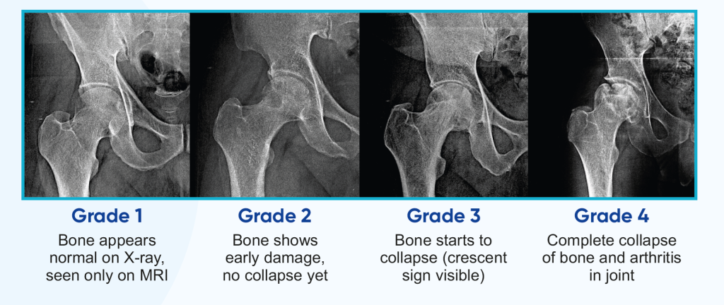
Treatment options shrink with every passing grade. At Grade 1–2, regenerative options like SVF therapy can help the bone heal. By Grade 4, hip replacement is usually the only reliable answer. The test you choose, and how soon you take it, directly decides which side of that line you land on.
MRI scan: the gold standard
MRI (Magnetic Resonance Imaging) uses magnets, not radiation, to photograph the inside of your bones. It is the most sensitive test for AVN and can detect the disease months or even years before an X-ray shows anything.
An MRI report for AVN typically describes:
- The extent of bone death, how much of the femoral head is affected
- Bone marrow edema, swelling inside the bone
- Fluid changes and early fracture lines invisible to X-rays
- Whether the other hip is silently affected too
Tip: Always ask for both hips to be scanned in the same sitting. AVN affects the second hip in a large share of patients, and catching it at Grade 1 is the best outcome possible.
X-ray imaging: useful, but late
X-rays are cheap, quick, and available everywhere in India. They show:
- The shape and structure of the femoral head
- Bone collapse or flattening (the "crescent sign")
- Joint space narrowing from arthritis
- Overall hip alignment
The catch: these changes appear only from Grade 2 onwards. A normal X-ray does not rule out AVN. If your X-ray is "fine" but the pain continues and you have risk factors, insist on an MRI.
Clinical examination
Before and alongside imaging, a specialist examines you in person:
- Range of motion testing: rotating and moving the hip to find restricted, painful directions
- Pain assessment: where it hurts, when, and how much
- Gait evaluation: watching how you walk, checking for a limp
- Medical history: steroid use, alcohol, injuries, other illnesses
- Risk factor identification: to protect the other hip and guide treatment
See the full picture of what AVN feels like on our symptoms page.
What to bring to your consultation
- Your MRI films or CD and the written report
- Any X-rays taken so far
- A list of medicines you take now or took in the past (especially steroids)
- Reports of related conditions, sickle cell, lupus, transplant, COVID hospitalisation
- A note of when the pain started and how it has changed
How we review your MRI: free, in 3 simple steps
- Send your details using the form below, or call +91 82230 82900.
- Share your MRI report when we call you back.
- Get a clear answer: your AVN grade, what it means, and which treatments genuinely fit your stage, explained in simple language, with no obligation.
Then read the complete AVN treatment guide to understand your options in depth.
Already Have an MRI or X-Ray? Get It Reviewed: Free.
Do not sit on a report you do not fully understand. We review your scans, confirm your AVN grade, and explain your real options, free and without obligation.
- Free MRI and X-ray report review
- Clear explanation in simple language
- In-person consultations at certified facilities
We do not provide telemedicine. All medical consultations happen in person at certified facilities.
Request a Call Back
Fill this in, it opens WhatsApp with your details ready to send.
AVN Diagnosis: Frequently Asked Questions
Can an X-ray alone diagnose AVN?
Only in later stages. In Grade 1 AVN the X-ray looks completely normal, because bone shape has not changed yet. A normal X-ray does not rule out AVN. If pain and risk factors point to AVN, an MRI is needed for a reliable answer.
How much does a hip MRI cost in India?
A hip MRI typically costs between ₹4,000 and ₹10,000 in most Indian cities, depending on the centre and machine strength. Compared to the cost of a delayed diagnosis, which can mean hip replacement, it is a very worthwhile investment. We can guide you to affordable, reliable centres near you.
Do I need contrast (dye) for an AVN MRI?
Usually not. A standard non-contrast MRI of both hips detects AVN reliably. Contrast is only used in special situations decided by the radiologist or specialist.
How often should I repeat an MRI if I have AVN?
Your specialist decides based on your grade and treatment. A common pattern is a follow-up MRI 3 to 6 months after starting treatment to check healing, and yearly checks afterwards. If pain suddenly worsens, an earlier scan may be needed.
Why should both hips be scanned?
AVN affects both hips in a large share of patients, especially steroid-related cases. Scanning both hips in one MRI sitting costs little extra and can catch the second hip at Grade 1, when it is easiest to save with joint-preserving treatment.
One Scan Can Save Your Hip.
If you have hip pain and risk factors, do not wait for an X-ray to "show something". Call us for a free consultation and get the right test at the right time.
Call for Free Consultation WhatsApp Us