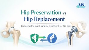
Understanding the stages of AVN is important because treatment decisions and outcomes depend heavily on how early the condition is diagnosed. While early-stage AVN may be managed with joint-preserving approaches, advanced stages often involve more complex treatment options.
Patients diagnosed in the early stages should explore treatment for AVN in India to better understand the available treatment options.
This guide explains all AVN stages (Stage 1 to Stage 4), how the disease progresses, the symptoms seen at each stage, and how treatment strategies change as AVN advances.
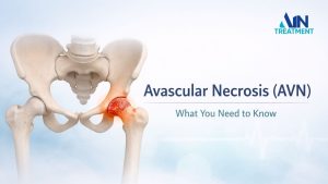
What Is Avascular Necrosis (AVN)?
Avascular necrosis is defined as death of bone tissue caused by reduced blood supply, as described in standard medical references. It is also called osteonecrosis.
Avascular necrosis is a condition in which bone tissue dies due to insufficient blood supply. Bone is a living tissue that constantly repairs itself. When blood flow is compromised for a prolonged period, bone cells begin to die, weakening the internal structure of the bone.
Read here to know what Avascular Necrosis (AVN) is.
Stages of AVN
MRI plays a critical role because early AVN stages may not appear on X-rays at all.
AVN is broadly divided into four stages, ranging from early blood flow disruption to complete joint collapse and arthritis.

Stage 1 (Early Stage):
Blood supply to the bone reduces, but the bone shape remains normal. Damage is microscopic and not visible on X-rays.
Most people have no symptoms. Some may feel mild, occasional discomfort in the hip or affected joint.
Monitoring, reducing joint stress, and managing risk factors. Early detection gives the best chance to slow progression.
Early detection at this stage offers the best chance to slow or halt progression.
Stage 2 (Pre-Collapse Stage):
Bone tissue damage increases, leading to structural weakness, but the femoral head has not yet collapsed.
Pain during walking or activity, reduced flexibility, and relief with rest.
Non-surgical, joint-preserving measures and load reduction. Timely treatment can help prevent collapse.
This stage is critical because timely intervention can prevent collapse.
Stage 3 (Collapse Stage):
Stage 3 marks a turning point in the disease. A subchondral fracture develops, and the femoral head begins to collapse.
Persistent pain, stiffness, difficulty walking or standing for long periods, and reduced range of motion.
Focus on preserving joint function, slowing further collapse, and improving quality of life. Advanced or surgical options may be considered.
At this stage, advanced and regenerative approaches may be considered, depending on patient factors and joint condition. Surgical options may also be discussed if symptoms progress.
Stage 4 (Advanced Stage):
Complete collapse of the femoral head with joint surface damage and secondary arthritis.
Severe, constant pain, marked stiffness, and major limitation in daily activities.
Pain relief and restoration of function. In many cases, surgical intervention becomes necessary due to extensive joint damage.
How Doctors Diagnose and Stage AVN
Imaging Techniques Used in AVN
- MRI (Magnetic Resonance Imaging):
The most sensitive test for detecting early AVN, even before symptoms appear. - X-ray:
Often normal in early stages; shows bone collapse in advanced stages. - CT Scan:
Occasionally used to assess bone structure in later stages.
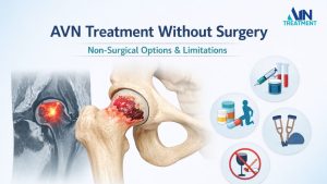
Can AVN Progression Be Slowed or Stopped?
AVN progression depends on:
- Stage at diagnosis
- Cause of AVN
- Timeliness of treatment
- Patient lifestyle factors
Early diagnosis and stage-appropriate care significantly improve outcomes. While advanced stages are harder to manage, appropriate treatment can still reduce pain and improve function.
Frequently Asked Questions About AVN Stages
Pain typically increases significantly in Stage 3 and Stage 4, when bone collapse occurs.
Early stages may be managed effectively, and progression can sometimes be slowed or halted.
Progression varies; some cases worsen over months, others over years.
No. Early and mid-stage AVN may be managed without surgery depending on individual factors.
Hip replacement is most commonly considered in Stage 4, when joint damage is severe.
Yes, AVN can also affect the knee, shoulder, ankle, and wrist, although the hip is the most commonly involved joint.
Not necessarily. With early diagnosis and appropriate management, progression may slow or stabilize in some patients.
Medical Disclaimer
This content is for educational purposes only and should not replace professional medical consultation. Diagnosis and treatment decisions should always be made by a qualified healthcare provider.

very helpfull