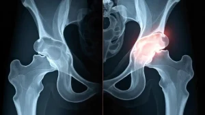
Avascular necrosis (AVN) of the hip is a condition that arises when the blood supply to the femoral head, the ball part of the hip joint, is reduced or completely cut off. This interruption causes the death of bone cells, leading to the weakening and eventual collapse of the hip joint, which impairs mobility and causes significant pain. In India, hip AVN often affects people between the ages of 30 and 50 and represents an important health issue due to lifestyle factors, steroid use, trauma, and infectious diseases like tuberculosis.
Detecting AVN early is critical because intervention at the initial stages can preserve the natural joint, delay or avoid the need for major surgery such as total hip replacement, and greatly improve the quality of life. This article outlines the early signs of hip AVN, how patients and medical professionals can recognise it sooner, the diagnostic methods involved, and treatment options tailored for early action.
Understanding Hip AVN and Its Importance
The hip joint depends on a delicate network of blood vessels to maintain the health and viability of the femoral head. When blood flow is blocked or diminished, the bone tissue starts to die, a process called avascular necrosis or osteonecrosis. Unlike many other bones in the body, the femoral head has a limited collateral blood supply, which makes it highly vulnerable to vascular interruption.
Without timely intervention, the bone weakens and collapses, causing deformity of the joint surface. As the disease advances, arthritis develops, and the patient experiences severe pain, stiffness, and loss of function. These factors underline the importance of recognising hip AVN at an early, often silent stage before irreversible damage occurs.
Causes and Risk Factors
In India, certain causes and risk factors significantly raise the likelihood of developing hip AVN. Trauma to the hip joint, such as fractures or dislocations, can directly damage the blood vessels supplying the femoral head. Long-term or high-dose corticosteroid use is another common cause. Steroids are widely used here for conditions like tuberculosis, asthma, and autoimmune diseases, all of which can unpredictably increase AVN risk.
Chronic alcohol use contributes to fat deposits blocking microcirculation, further impairing blood flow. Blood disorders, particularly sickle cell disease or thrombophilia, may compromise vascular supply as well. Other causes include radiation therapy, decompression sickness (common among divers), and idiopathic origins.
Recognising Early Symptoms: Subtle Clues to Hip AVN
Early symptoms of hip AVN often present as mild and nonspecific, leading to missed or delayed diagnosis. The most common initial complaint is a gradual onset of dull, aching pain located in the groin area. Patients often report pain intensifying during activities that load the hip, such as walking, climbing stairs, or standing for extended periods. The pain might radiate to the thigh or buttocks, sometimes confusing the source.
In the initial stages, the discomfort may occur only during movement but can later develop into rest pain, disturbing sleep, and reducing overall comfort. Stiffness in the hip joint emerges, particularly restricting internal rotation and abduction movements.
Patients may notice difficulty sitting cross-legged or squatting positions commonly used in Indian culture, thus affecting daily routines.
A subtle limp or altered gait pattern frequently develops as individuals subconsciously protect the painful joint.
This slow, progressive character of symptoms often leads patients to attribute the pain to minor strain or arthritis.
Hence, awareness of these early, often overlooked signs is essential for both patients and healthcare providers.
Clinical Examination and Its Role in Early Detection
A thorough clinical examination provides valuable clues that support early detection of hip AVN. The healthcare provider will test the hip’s range of motion and may elicit pain on passive and active movement, particularly with internal rotation or abduction. Tenderness over the groin area or just below the greater trochanter (the prominent bone on the side of the hip) may also be present.
In early disease, muscle strength around the hip may still be preserved, but as AVN progresses, signs of weakness can be detected. The Trendelenburg test, which assesses hip abductor function by observing pelvic tilt during single-leg stance, may become positive in later stages but is still potentially manageable.
While physical examination alone cannot confirm AVN, these findings combined with risk factor assessment should raise suspicion, prompting further investigation.
Diagnostic Imaging: Confirming the Diagnosis Early
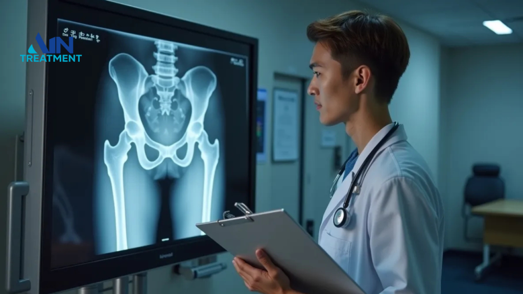
Imaging plays a pivotal role in confirming AVN and its stage. Standard X-rays are often the initial test due to their wide availability and low cost, but they lack sensitivity in early disease. In the initial stages of AVN, X-rays typically appear normal because structural changes have yet to develop.
Magnetic Resonance Imaging (MRI) is the most sensitive and specific modality for early detection. MRI identifies changes in bone marrow composition, presence of oedema, and necrotic areas before X-ray evidence of disease appears. This allows diagnosis in the earliest stages (Stage I or II by common classification systems), when joint-preserving treatments are most effective.
Other diagnostic tools, such as bone scans, offer information on blood flow deficits, but their specificity is lower. Computed tomography (CT) scans provide excellent detail of bone architecture and are useful when assessing collapse or deformity at later stages, but are less helpful early on.
Staging of Hip AVN and Its Clinical Relevance
Staging helps guide treatment decisions. Early stages (I and II) are characterised by no visible collapse of the femoral head but some changes visible on MRI and sometimes subtle X-ray changes. Stage III includes evidence of femoral head collapse without joint space narrowing, while Stage IV represents advanced collapse with secondary arthritis.
Correct staging is essential because early, non-surgical and minimally invasive treatments are effective primarily in stages I and II. Once collapse and arthritis develop, joint-preserving options become ineffective, and replacement surgery becomes necessary.
Early Intervention: What Can Be Done?
Lifestyle changes often form the first step. Patients are advised to limit weight-bearing activities and use aids like walking sticks or crutches to reduce stress on the femoral head.
Medications such as bisphosphonates might reduce bone resorption and improve bone strength, although evidence varies.
Pain relief through analgesics and anti-inflammatory drugs enhances comfort but does not address the underlying cause. Physiotherapy focuses on maintaining muscle strength around the hip and maintaining joint mobility.
Among surgical options, core decompression is the most frequently employed in early AVN.
This involves removing a small portion of inner bone from the femoral head and neck to relieve intraosseous pressure, promote new blood vessel growth, and stimulate repair. Core decompression has the best success rate when performed before collapse occurs.
In some cases, bone grafting may be performed alongside core decompression to fill the defect and encourage bone regeneration. Osteotomy, involving the realignment of bone to offload the damaged area, is another option, but technically demanding.
Innovative treatments like stem cell therapy are gaining traction in India and worldwide, aimed at regenerating bone through biological means; however, their role is still being studied.
When Does Surgery Become Inevitable?
If AVN progresses despite early interventions, femoral head collapse ensues, and secondary osteoarthritis develops. At this stage, joint-preserving treatments fail to provide relief or restore function. Total hip replacement (THR) becomes the definitive treatment option.
While highly successful for pain relief and restoring mobility, THR has limitations, especially in young active patients typical of the Indian population. Prostheses have a limited lifespan, and activity restrictions post-surgery impact quality of life, which underscores the importance of early detection and intervention.
Monitoring and Follow-Up

Regular follow-up of patients with risk factors or early symptoms is critical. Periodic clinical assessments allow early recognition of symptom progression. Imaging, primarily MRI, should be repeated as necessary to monitor disease progression and response to treatment.
Educating patients about symptoms and the importance of early consultation enhances timely diagnosis. Healthcare providers across primary and secondary care levels should maintain a high index of suspicion in appropriate cases.
Challenges
Several challenges complicate the early detection of hip AVN . Patients often delay seeking medical help due to a lack of awareness or access issues, especially in rural areas where MRI and specialist consultations may be unavailable. There is frequent misdiagnosis as simple arthritis or muscular strain.
Widespread corticosteroid use without adequate monitoring is a significant concern. Moreover, lifestyle and occupational factors such as prolonged squatting or heavy manual labour can exacerbate symptoms and delay diagnosis.
Addressing these challenges requires public health education, better primary care training, and improved access to diagnostic facilities.
Conclusion
Hip AVN is a progressive condition that demands early recognition and timely intervention to protect the natural joint and prevent disability. Early signs such as mild groin pain, activity-related discomfort, and subtle stiffness should prompt evaluation, especially in patients with known risk factors. Although X-rays are standard first investigations, MRI remains the gold standard for early detection.
